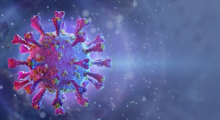
In a recent study published in Cells, researchers investigated coronavirus disease 2019 (COVID-19) pathophysiology in multiple tissues, including the lungs, kidneys, heart, and brain, for a deeper understanding of its acute and long‐term symptoms.
Study: COVID-19 Pathology within the Lung, Kidney, Hearts and Brain: The Different Roles of T-Cells, Macrophages, and Microthrombosis. Image Credit: Corona Borealis Studio/Shutterstock
Background
Coronaviruses (CoVs), including severe acute respiratory syndrome coronavirus 2 (SARS-CoV-2), typically infect the respiratory and digestive tracts of host species. Nonetheless, pathological studies have confirmed that SARS-CoV-2 infection clinically manifests in multiple organs. The extent of the involvement of various organs might be a reliable marker of COVID-19 hostile outcomes and PASC. PASC, i.e., ongoing symptomatic COVID‐19 beyond 12 weeks, may affect the hosts’ lungs, kidneys, heart, and brain with variable severity.
Concerning the study
In the current study, researchers performed a radical cross-sectional evaluation of clinical and pathological data from COVID‐19 cases from the primary pandemic peak. Moreover, they evaluated this data for the presence of Alzheimer’s’ disease (AD) neuropathology. They chose nine of 15 COVID‐19 autopsies for the study evaluation performed between April 17 and June 4 2020. There have been 10 controls, of which five cases had non‐COVID-19 pneumonia. The remaining five had non‐COVID-19 brains, including three AD cases.
Two forensic medical doctors, a geriatrician, and a neurologist performed a retrospective review of the medical charts of all nine study participants. They clinically ascertained the presence or absence of comorbidities, dementia, delirium, and sepsis. The team harvested the tissue samples from SARS-CoV-2-infected cadavers for pathological examinations.
It included one section per pulmonary lobe, five heart sections from a mid‐horizontal slice, and one section from each kidney, including the cortex and medulla. Additionally they harvested seven brain sections from the frontal, temporal, parietal, and occipital lobes; hippocampus–entorhinal cortex; pons; and cerebellum. The team also performed the routine histological examination of those sections using the hematoxylin-eosin (H&E) staining technique.
Moreover, the researchers quantified SARS-CoV-2 ribonucleic acid (RNA) load in these tissues via quantitative reverse‐transcription–polymerase chain response (qRT‐PCR) and droplet digital PCR (ddPCR). Finally, the researchers checked inflammatory infiltrates and microthrombi within the inferior left lung lobe, right kidney, left heart ventricle, frontal lobe for the forebrain, and pons for the hindbrain, for the presence of SARS-CoV-2. Because the variety of cases was low, the team used a non-parametric statistical test and Friedman’s test to match different tissues inside subjects. They used the Mann–Whitney U test to match rating differences between the cases and controls for every organ.
Study findings
The first study finding was that persistent inflammation and never persistent SARS-CoV-2 replication were more than likely liable for the PASC. Inflammatory and immune‐mediated alterations augment the direct cytopathic damage triggered by SARS-CoV-2. These accentuate epithelial and endothelial damage, vascular leakage, and dampening of the T‐cell response, together with the over‐activation of the macrophage lineage cells; hence, it encompasses multiple organs. In some cases, lung failure causes severe hypoxia in all tissues.
The authors found that the organ with the heaviest burden of SARS-CoV-2 antigens was the lung, and kidneys shared the burden, but to a lesser extent. Nonetheless, the guts and brain had rare endothelial cells or pontine neurons and absolutely no evidence of energetic SARS-CoV-2 replication. In comparison with controls, the COVID-19 deceased patient’s lungs also had markedly higher microthrombosis and abundant T‐lymphocytes.
Moreover, they observed that the guts had rare T-lymphocytes, minimal SARS‐CoV‐2 traces within the endothelium–endocardium, and foci of activated macrophages. The brain similarly showed minimal traces of SARS‐CoV‐2 and rare T-lymphocytes but outstanding microglial activation within the frontal cortex that was probably related to pre‐existing neurodegeneration on account of AD relatively than COVID‐19. Quite the opposite, the pons exhibited the best microglial activation related to SARS‐CoV‐2 infection.
Conclusions
The present study presented a remarkable pathological picture of how SARS-CoV-2 invades various human tissues. It demonstrated that lung and kidney tissue damage may be directly on account of SARS-CoV-2-induced cytopathic effects and subsequent inflammation‐mediated mechanisms. Nonetheless,
COVID-19-induced damage to the guts and brain was mainly on account of aberrant and chronic inflammation. Thus, a whole recovery from COVID‐19 requires the termination of each, which might take many months.
A deeper understanding of the short- and long-term detrimental effects of COVID-19 could help improve look after COVID‐19 patients, especially after the acute phase of the disease. As an example, some patients might require physical and cognitive training and psychosocial support on priority during recovery to revive previous functionalities.
Journal reference:
- COVID‐19 Pathology within the Lung, Kidney, Hearts and Brain: The Different Roles of T‐Cells, Macrophages, and Microthrombosis, Tino Emanuele Poloni, Matteo Moretti, Valentina Medici, Elvira Turturici, Giacomo Belli, Elena Cavriani, Silvia Damiana Visonà, Michele Rossi, Valentina Fantini, Riccardo Rocco Ferrari, Arenn Faye Carlos, Stella Gagliardi, Livio Tronconi, Antonio Guaita, Mauro Ceron. Cells. doi: https://doi.org/10.3390/cells11193124 https://www.mdpi.com/2073-4409/11/19/3124