In a recent study posted to the bioRxiv* preprint server, researchers investigated severe acute respiratory syndrome coronavirus 2 (SARS-CoV-2) envelope (E) protein activity by way of calcium cations (Ca2+) cations.
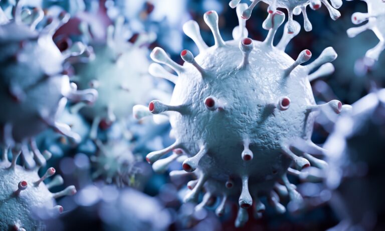
Study: The SARS-CoV-2 envelope (E) protein forms a calcium- and voltage-activated calcium channel. Image Credit: PHOTOCREO Michal Bednarek/Shutterstock
Functional ion channels are critical within the infectious cycles of several viruses since viruses modify host ionic balance (especially Ca2+) to facilitate their uptake, maturation, and export. Viroporins encoded in viral genomes are essential for altering ionic and cellular homeostasis. SARS-CoV-2 E forms ion channels within the endoplasmic reticulum (ER)-Golgi intermediate compartment (ERGIC) membranes in association with SARS-CoV-2 virulence and progression of infection.
Studies have reported that blockade, deletion, or loss-of-function mutations in CoV E proteins can generate attenuated or propagation-lacking viral variants; nonetheless, precise physiological functions of SARS-CoV-2 E are usually not well-characterized and require further investigations.
Concerning the study
In the current study, researchers explored the prime physiological function of SARS-CoV-2 E upon viral infection.
E protein construct comprising the full-length E sequence or residues 1 to 75 (EFL) was produced, purified from E. coli inclusion bodies, and reconstituted into phosphatidylethanolamine (PE) membranes under voltage-clamp conditions. EFL oligomers were formed and confirmed by Western blot evaluation and mass photometry (MP).
Molecular dynamic (MD) simulations were performed, and voltage-clamp electrophysiological measurements were recorded to quantify Ca2+ channel activity. The membrane-bound structure and functional ion channel activities of SARS-CoV-2 E were investigated. EFL pentamerization was performed and confirmed by size exclusion chromatography coupled with multi-angle light scattering (SEC-MALS) evaluation.
As well as, the consequences of post-translational modifications (PTM) on the E protein function were explored by palmitoylating all of the cysteine residues (Cys40, Cys43, Cys44) in every subunit within the EFL pentamers of SARS-CoV-2 E protein channels. Further, the consequences of luminal Ca2+ concentrations on EFL gating properties were evaluated.
The team investigated if the transmembrane (TM) site formed EFL functional substructures, for which ETM was produced comprising viral E protein residues 8 to 38, by solid-phase peptide synthesis and assessed ETM functionality in-vitro. The team investigated whether ETM was inserted into PE planar lipid bilayers under voltage-clamp conditions and performed MD simulations on ETM domains within the assembled pentamers.
Results
SARS-CoV-2 E formed Ca2+-permeable ion channels within the planar lipid bilayers, which relied on hydrophobic gating and lipids. The viral E protein exhibited a binding annulus for Ca2+ ions at the doorway of the luminal pores that stabilized the pores in open states. In consequence, calcium cations increased open durations of the pores and ionic currents passing through the E protein ion channels.
The hydrophobically gated ion channel activity of the viral E protein and viroporins were regulated by elevated luminal Ca2+ concentrations (0.1 mM to 1.0 mM), electrochemical gradients, pH, PTMs, ERGIC phospholipids with negative charges, and voltage applied to the membranes. Palmitoylation of ≥1 cysteine residue promoted the formation of open and stable E protein pores. Ca2+ ions activated ER-luminal channels and maintained the pores within the open state.
Ca2+-Glutamic residue interactions altered E protein conformation and favored ion channel opening and the flow of ions into and thru the channels. The distinctive calcium-binding site within the E channels served as a recruitment region for ions and an activation site within the pores. SEC-MALS and MP evaluation findings showed that EFL pentamers were the prevailing states of the E protein construct. The E protein showed cation selectivity over anions, with Cl- permeability one-third of Na+ permeability.
By utilizing Ca2+ because the permeant cation, the team observed multiple channel incorporation and frequent but temporary open events to several open states and better permeability of viroporin to Na+ than Ca2+ ions. The voltage experiments showed that the E protein was almost certainly a voltage-gated pore regulated by electrowetting and a hydrophobic gating motif (comprising Phe20, 23, and 26 residues) positioned within the pore’s center.
The TM domain, individually, didn’t form physiologically functional substructures of the viral E protein. Due to this fact, the domain constructs is probably not appropriate models to realize insights into the viral E protein function and structure for developing anti-SARS-CoV-2 drugs. Ca2+ release via the viral E protein pore depended strongly on Ca2+ loads and Ca2+ store depletion below threshold or the positive ion binding region modulation could abolish SARS-CoV-2 E-mediated Ca2+ flux. The finding is critical, given evidence of calcium ion dysregulation in cells in coronavirus disease 2019 (COVID-19).
Overall, the study findings highlighted the physiological role of SARS-CoV-2 E involving Ca2+ release from the ER and that the distinctive Ca2+ activation region might be potentially targeted for the event of anti-CoV agents based on ion channel blockage mechanisms. The findings highlighted novel ion and lipid interaction sites on SARS-CoV-2 E that might be targeted for developing anti-SARS-CoV-2 drugs, potentially stopping fatal excess stimulation of host immune responses and addressing the least amino acid substitution-prone a part of the SARS-CoV-2 proteome.
*Vital notice
bioRxivpublishes preliminary scientific reports that are usually not peer-reviewed and, due to this fact, mustn’t be considered conclusive, guide clinical practice/health-related behavior, or treated as established information.