In a recent study posted to the bioRxiv* preprint server, researchers identified β-cyclodextrins (β-CDs) as a protected and cost-effective drug against severe acute respiratory syndrome coronavirus 2 (SARS-CoV-2) with a broad spectrum of activity.
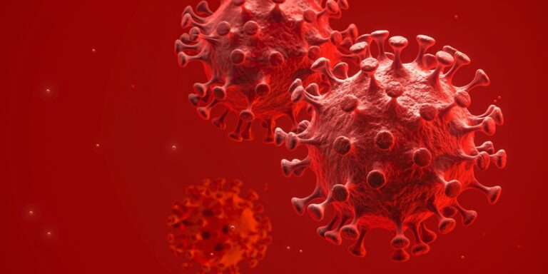
Study: β-Cyclodextrins as reasonably priced antivirals to treat coronavirus infection. Image Credit: 3DJustincase/Shutterstock
COVID-19 (coronavirus disease 2019) has led to significant global morbidity and mortality. No anti-SARS-CoV-2 agent authorized to be used is economical, easy to make use of, or can provide global COVID-19 prophylaxis for people at high risk of disease severity outcomes. The event of economic, broad-spectrum, and protected therapeutics on the onset of novel SARS-CoV-2 variant emergence could curtail transmission of SARS-CoV-2 to a substantial extent.
In regards to the study
In the current study, researchers identified β-cyclodextrins (β-CDs) as cost-effective and broad-spectrum COVID-19 therapeutic agents protected for human administration.
The team enlisted 116 drugs which were used previously for treating pathologies or have demonstrated anti-SARS-CoV-2 efficacy in pre-clinical trials. Molecular modeling was performed to rank 44 compounds that showed the best efficacy against several α CoVs and β CoVs, e.g., SARS-CoV-2 and human CoV 229E (HCoV-229E). The drugs were identified by virology, computational chemistry, transmission electron microscopy (TEM), and cell biology techniques.
The antiviral mechanisms of the compounds were explored by TEM findings and the flexibility to inhibit SARS-CoV-2 pseudoviruses’ entry into angiotensin-converting enzyme 2 (ACE2)-expressing human embryonic kidney (HEK)-293T cells. Protein data bank (PDB) structures underwent refinement, states of protonation were determined, restricted minimization was performed, and grids were created. Further, six native and modified cyclodextrins were tested.
Binding energies were computed based on the molecular mechanics/generalized Born surface area (MMGBSA) evaluation, the outcomes of which were analyzed to rank the compounds based on their antiviral efficacy. Further, molecular dynamics (MD) simulations were performed to evaluate the steadiness of the compounds. HCoV-229E was propagated in MRC-5 cells (medical research council cell strain 5), following which, the cells were subjected to indirect immunofluorescence (IF) evaluation using anti-HCoV-229E nucleocapsid (N) protein antibodies.
Moreover, MTT (3-[4,5-dimethylthiazol-2-yl]-2,5 diphenyl tetrazolium bromide) assays for cell viability assessments and the antiviral efficacy of the identified compounds were evaluated based on IF findings. SARS-CoV-2 variants whose genomic sequences were uploaded within the GISAID (global initiative on sharing all influenza data) database were isolated from nasopharyngeal swab samples in Vero E6 cells, and the supernatant was subjected to genomic sequencing.
SARS-CoV-2 spike (S) protein-expressing human immunodeficiency virus 1 (HIV-1) reporter pseudoviruses were generated. Luminometric assays and transfection experiments were performed. The p24gag content of viruses was measured using enzyme-linked immunosorbent assays (ELISA), following which the viruses were titrated in ACE2-overexpressing HEK-293T cells. As well as, pseudoviral entry inhibition assays were performed, and SARS-CoV-2 replication was assessed using Calu-3 cells (human lung cancer cell line). SARS-CoV-2 foremost protease (Mpro) inhibition assays and the cell membranes of Calu-3 cells were subjected to lipidomic evaluation post-MbCD (methyl-b-CD) treatment.
Results
4 compounds, U18666A, OSW-1, phytol, and hydroxypropyl-β-cyclodextrin (HβCD) demonstrated antiviral activity against HCoV-229E and SARS-CoV-2 in MRC-5 cells and Vero E6 cells, respectively. U18666A and HβCD blocked viral fusion; nonetheless, only HβCD showed inhibition of SARS-CoV-2 replication in Calu-3 cells. Testing native and modified cyclodextrins confirmed the potent SARS-CoV-2 inhibition by β-cyclodextrins in Calu-3 cells.
OSW-1 anchored efficiently on the lively Mpro site with the biggest binding energy amongst all molecules tested. b-CD and OSW-1 localized within the catalytic pocket of Mpro, whereas phytol and U1866A localized within the central pocket of NPC1. Each OSW-1 and b−CD were stable with mean RMSD (root means-square deviation) variations of ca. 2Å and the values were double for phytol and U18666A.
The half maximal inhibitory concentration (IC50) values for U18666A, OSW-1, HβCD, and phytol and were 2.6µM, 0.5nM, 4.3mM, and 19µM, respectively. Cyclodextrins inhibited SARS-CoV-2 replication by interfering with viral fusion via cholesterol depletion. HβCDs, MbCDs, and b-CDs inhibited SARS-CoV-2 D614G strain and BA.1 strain on Calu-3 cells.
TEM findings of SARS-CoV-2-infected Vero E6 cells showed that U18666A and OSW-1 affect SARS-CoV-2 morphogenesis by inhibiting double-membrane vesicle (DMV) assembly and performance. U18666A high-dose treatment led to the enlargement of lysosomes and Golgi cisternae. Phytol low-dose treatments altered DMVs to a limited extent with a minor impact on SARS-CoV-2 assembly and transmission, whereas clear cytopathic effects were visualized using high-dose phytol.
HβCD treatment at 0.2 mM concentration altered the inner membrane of DMVs, with large clusters of distorted SARS-CoV-2 particles observed inside the vacuoles. At 20 mM HβCD concentration, all structures of SARS-CoV-2 were reduced with few altered DMVs.
Overall, the study findings highlighted β-cyclodextrins as promising therapeutic agents against SARS-CoV-2 and potentially other pulmonary viral organisms.
*Necessary notice
bioRxiv publishes preliminary scientific reports that usually are not peer-reviewed and, subsequently, mustn’t be thought to be conclusive, guide clinical practice/health-related behavior, or treated as established information.